
3D Cone Beam Imaging in Summerville, SC
A blurry two-dimensional X-ray can only tell part of the story when something feels wrong beneath the surface of a tooth or jaw. If you have ever been told you need “more imaging” before a procedure can move forward, you know how frustrating it feels to wait on answers about your own mouth.
At Old Trolley Dental Associates, we use 3D cone beam imaging to see what flat X-rays cannot. This technology captures a complete three-dimensional view of your teeth, jawbone, sinuses, and nerve pathways in a single scan, giving us a clearer picture before we ever discuss dental implants or other advanced treatment.
What Is 3D Cone Beam Imaging?
Cone beam computed tomography, often shortened to CBCT, is a specialized form of dental X-ray technology that rotates around your head to build a detailed 3D model of your oral anatomy. Instead of a single flat image, we get hundreds of individual snapshots that combine into one comprehensive view.
This means we can examine bone density, root positions, nerve locations, and airway structure from any angle we need. A traditional X-ray might hint that something is off, but a cone beam scan lets us confirm it and plan around it with precision.
Why Precision Imaging Matters
Every mouth is shaped differently, and guesswork has no place in a treatment plan. Research published through the National Center for Biotechnology Information has examined how CBCT dose settings can be adjusted across different clinical indications while maintaining diagnostic image quality, which speaks to how carefully this technology has been studied and refined for patient safety.
We rely on this level of detail most often when planning dental implant placement, since knowing the exact width and height of your jawbone helps us select the right implant size and position before any drilling begins. The same imaging also helps us evaluate impacted teeth, plan complex extractions, and identify issues that would otherwise go unnoticed until a problem became painful.
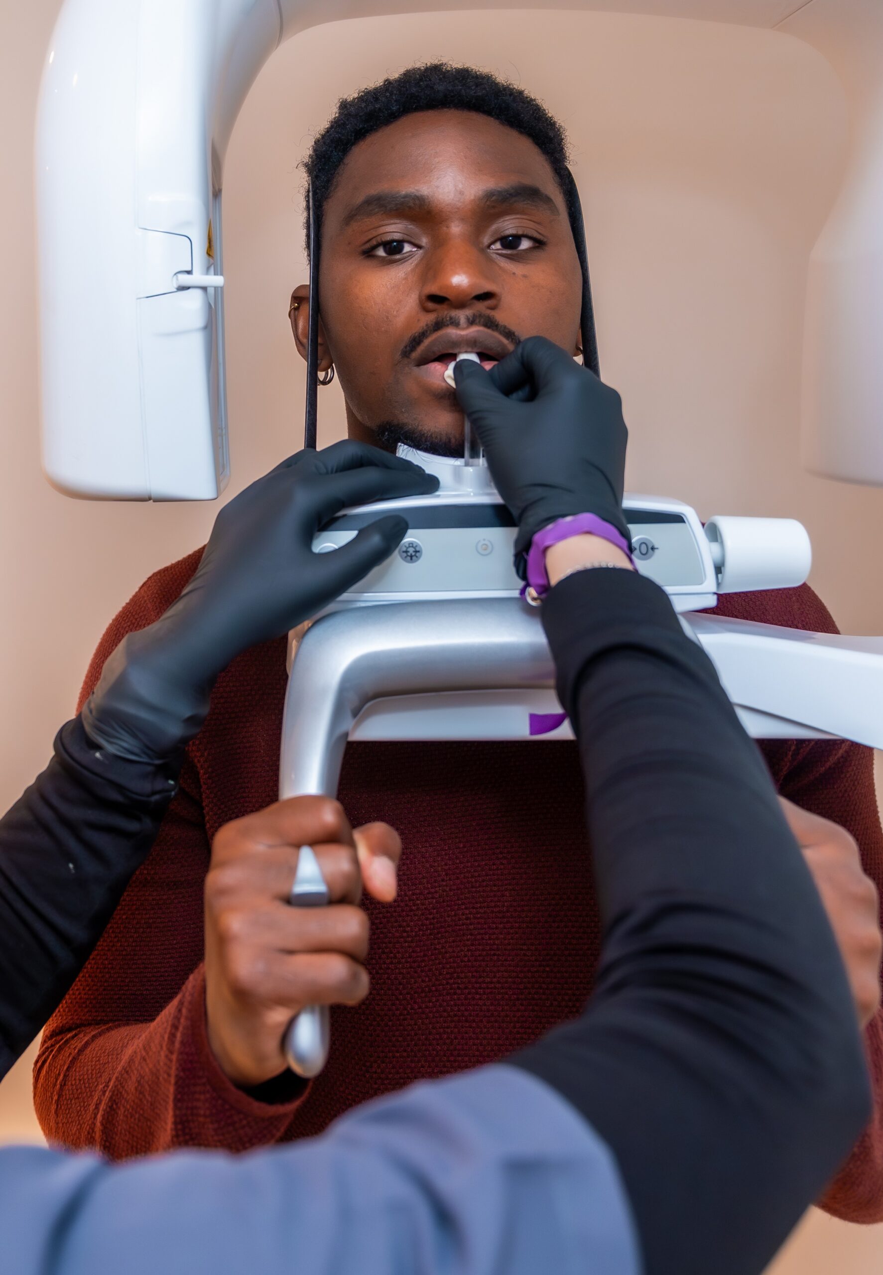
How We Use Cone Beam Imaging in Your Care
A cone beam scan takes only a few seconds and requires you to simply sit still while the machine rotates around your head. There is no biting down on uncomfortable film or holding an awkward position, and you can see your results with us the same day.
We use this imaging to guide several types of treatment planning, and having this level of detail up front tends to make the rest of your treatment more predictable. Some of the most common reasons we recommend a cone beam scan include the following:
- Mapping bone structure and nerve position before dental implant placement
- Evaluating impacted or difficult-to-reach wisdom teeth before extraction
- Identifying hidden infections or unusual root anatomy during root canal therapy
- Assessing jaw joint health, airway space, or sinus involvement related to a treatment plan
Once we have reviewed your scan, we walk you through exactly what we see and what it means for your treatment options. This conversation is where the real value of the imaging shows up, since a picture only helps if you understand what it is telling us about your specific situation.
What to Expect During Your Scan
You will not need to do anything special to prepare for a cone beam scan. We ask you to remove any metal jewelry or eyewear that could interfere with the image, and then you will sit or stand in the machine for a brief rotation that typically takes less than 30 seconds.
Because the scan is so quick and comfortable, most patients are surprised by how simple the process feels compared to what they expected. If your treatment involves wisdom teeth removal or another procedure where positioning matters, this imaging often becomes part of your very first visit so we can start planning right away.
Cone Beam Imaging Supports More Confident Treatment Decisions
Diagnostic clarity changes the way treatment gets planned. When we can see the exact shape of your jawbone, the path of a nerve, or the anatomy of a root canal system before we start, we can explain our reasoning to you in a way that makes sense and avoid surprises once treatment is underway.
This technology also supports more conservative treatment in many cases. Seeing precisely where a nerve sits or how much healthy bone is available may allow us to choose a less invasive approach than we would have selected without that information, which can mean a smoother recovery and fewer follow-up visits.
Schedule Your Visit With Old Trolley Dental Associates
Old Trolley Dental Associates has served Summerville families since long before our current name, growing from a Navy veteran’s practice into the two-doctor team led by Dr. Scott Kibler and Dr. Michael Skovira today. Our office combines advanced diagnostic tools like 3D cone beam imaging with same-day crown technology and Dr. Skovira’s background in digital smile design, giving patients access to precise, modern care without long waits between appointments or referrals to outside specialists.
We accept a range of PPO insurance plans and offer Cherry and CareCredit financing to help make treatment planning easier on your budget. If you would like to learn more about how our doctors use cone beam imaging in your treatment plan, contact our office today to schedule your visit.